In a Time of Change
collected by the Anchorage Museum
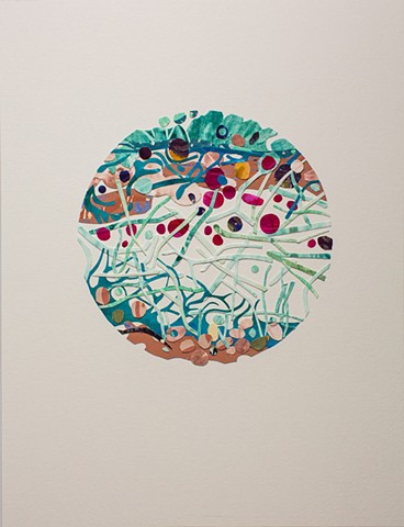
Lichen Cross Section is an observational piece inspired by the cellular structure of a lichen. Lichen are composed of algal or cyanobacterial cells living symbiotically with strands of fungi. It was recently discovered that yeast, which are another type of fungi, are a third partner in the lichen symbiosis. Lichen are interesting in that we can see them without the use of a microscope. The many algal and fungal components are microscopic, yet when they are all together they make up the plant-like organisms we see on rocks and branches. This piece zooms in to show the lichen under a microscope.
I viewed different types of lichen in a variety of ways. Initially, I swabbed the surface and cultured agar plates. This revealed a wealth of life, but not the cellular structure I was looking for. Preparing slides with feather-thin slices of lichen provided me with the visually and metaphorically rich imagery I craved. I took photographs through the lens of the microscope and used the photographs along with scientific illustrations of lichen cross-sections to create this piece. The forms and colors are based on biology but are infused with imagination.
The processes of science and art are so similar. With both, a question or problem needs to be solved and the path to solve it is rarely straight. I found that preparing slides and using a microscope required skills and demanded practice much like using a printing press or mixing colors.